fbpx Skip to main content

Spine Center Services

dekomp_1280

Discectomy

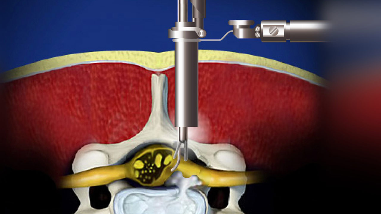
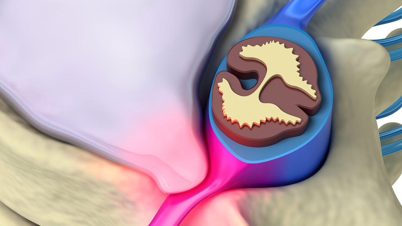
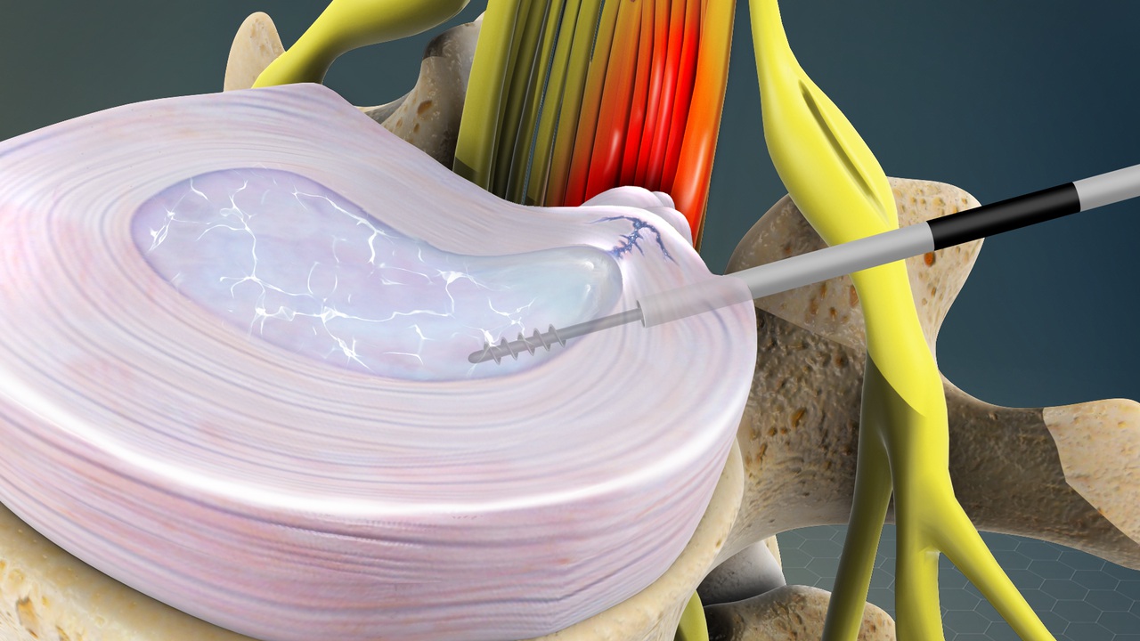
Discectomy is surgery to remove lumbar (low back) herniated disc material that is pressing on a nerve root or the spinal cord. It tends to be done as a microdiscectomy, which uses a special microscope to view the disc and nerves. This larger view allows the surgeon to use a smaller cut (incision). And this causes less damage to surrounding tissue.

Before the disc material is removed, a small piece of bone (the lamina) from the affected vertebra may be removed. This is called a laminotomy or laminectomy. It allows the surgeon to better see the herniated disc. Discectomy is usually done in a hospital. You are asleep or numb during the surgery. You will probably stay in the hospital overnight.

Schedule a Consultation

Microdiskectomies

Spinal surgery once meant large incisions, long recovery periods, and painful rehab. Fortunately, surgical advances like the microdiscectomy procedure have improved the process.

Microdiscectomy, also sometimes called microdecompression or microdiscectomy, is a minimally invasive surgical procedure performed on patients with a herniated lumbar disc. During this surgery, a surgeon will remove portions of the herniated disc to relieve pressure on the spinal nerve column.

Schedule a Consultation
Microdiskectomies
Laminectomy

Laminectomy with Fusion or Instrumentation

Laminectomy is surgery that creates space by removing the lamina — the back part of a vertebra that covers your spinal canal. Also known as decompression surgery, laminectomy enlarges your spinal canal to relieve pressure on the spinal cord or nerves. This pressure is most commonly caused by bony overgrowths within the spinal canal, which can occur in people who have arthritis in their spines.

These overgrowths are sometimes referred to as bone spurs, but they’re a normal side effect of the aging process in some people. Laminectomy is generally used only when more conservative treatments — such as medication, physical therapy, or injections — have failed to relieve symptoms. Laminectomy may also be recommended if symptoms are severe or worsening dramatically.

Schedule a Consultation

Kyphoplasty

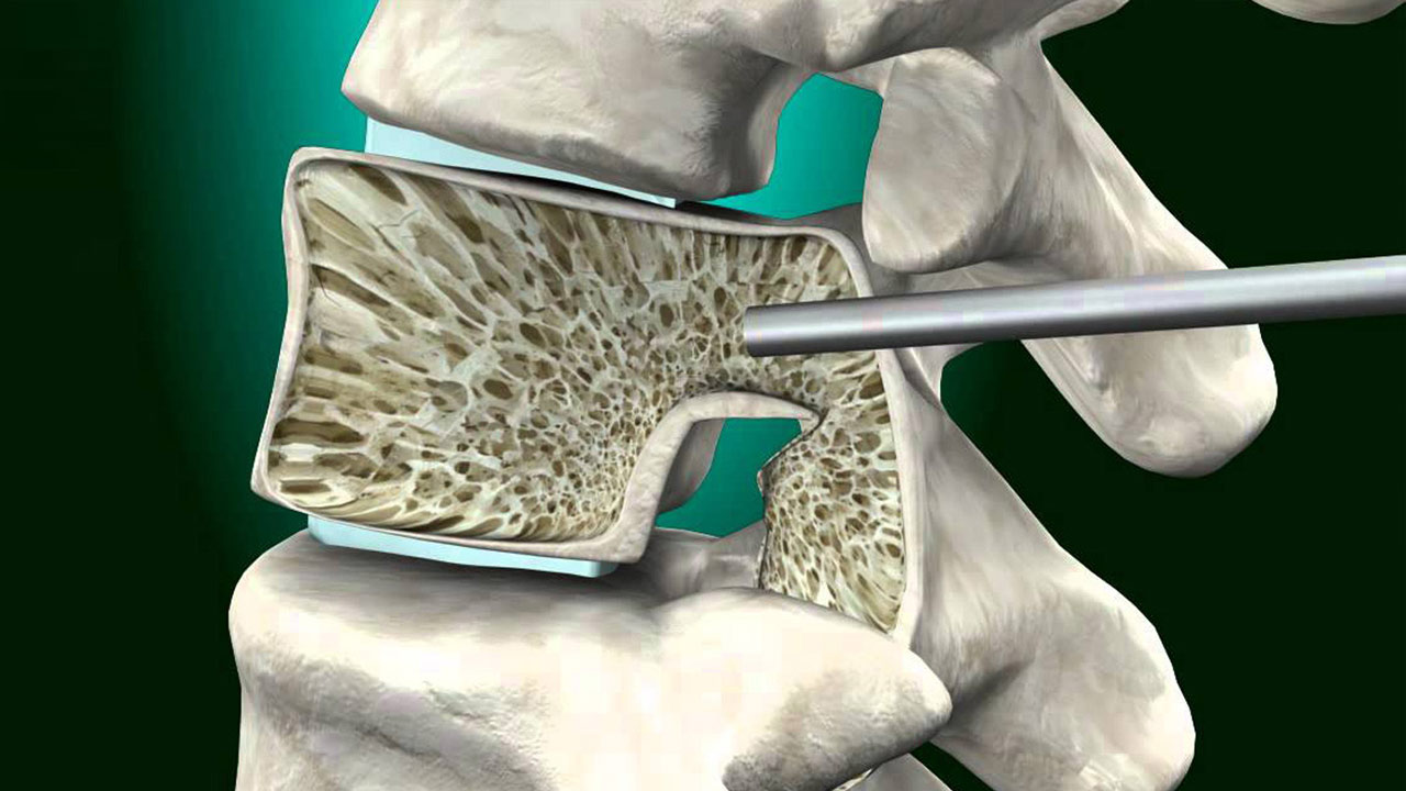
Vertebroplasty and kyphoplasty are minimally invasive procedures for the treatment of painful vertebral compression fractures (VCF), which are fractures involving the vertebral bodies that make up the spinal column. When a vertebral body fractures, the usual rectangular shape of the bone becomes compressed, causing pain. These compression fractures may involve the collapse of one or more vertebrae in the spine and are a common result of osteoporosis.

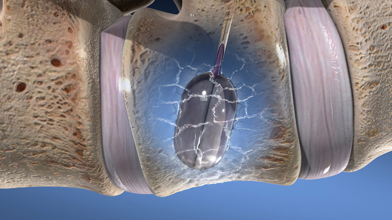
Osteoporosis is a disease that results in a loss of normal bone density, mass, and strength, leading to a condition in which bones become increasingly porous and vulnerable to breaking easily. Vertebrae may also become weakened by cancer. For vertebroplasty, physicians use image guidance, typically fluoroscopy, to inject a cement mixture into the fractured bone through a hollow needle. During kyphoplasty, a balloon is first inserted into the fractured bone through the hollow needle to create a cavity or space. The cement is injected into the cavity once the balloon is removed.

Schedule a Consultation
Laminectomy
Kyphoplasty

Vertebroplasty

Vertebroplasty is an outpatient procedure for stabilizing compression fractures in the spine. Bone cement is injected into back bones (vertebrae) that have cracked or broken, often because of osteoporosis. The cement hardens, stabilizing the fractures and supporting your spine.

For people with severe, disabling pain caused by a compression fracture, vertebroplasty can relieve pain, increase mobility, and reduce the use of pain medication. Kyphoplasty is similar to vertebroplasty but uses special balloons to create spaces within the vertebra that are then filled with bone cement. Kyphoplasty can correct spinal deformity and restore lost height.

Schedule a Consultation

Cervical and Lumbar Disk Replacement

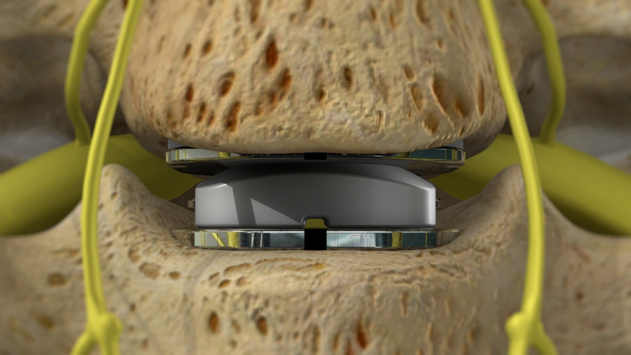
Cervical disc replacement is a surgical procedure that involves removing a damaged or degenerated cervical disc and replacing it with an artificial disc device. Cervical discs are the cushions or shock absorbers between the bones (vertebra) of the neck (cervical spine). Discs that become damaged either through trauma or degeneration can be a source of pain.

If part of a disc moves out of its normal position it can cause pressure on the central spinal cord or on the individual nerve roots that exit from the spinal canal at each vertebral level.

Schedule a Consultation
Spine Center Services

Revision Spine Surgery

Revision spinal surgery is a procedure that takes place on a patient who has already undergone some form of spine surgery. Typically, by three months after a surgery has been completed, any lingering pain is gone. When a patient is still reporting chronic pain beyond this time frame, revision spinal surgery is sometimes considered.

Revision spinal surgery may also be related to the herniation of a disc, infection, hardware failure, non-surgery related spine degeneration, flat back syndrome, instability, adjacent segment degeneration, or pseudarthrosis (failure to achieve solid fusion).

Schedule a Consultation

Lumbar Percutaneous Fusion and Instrumentation

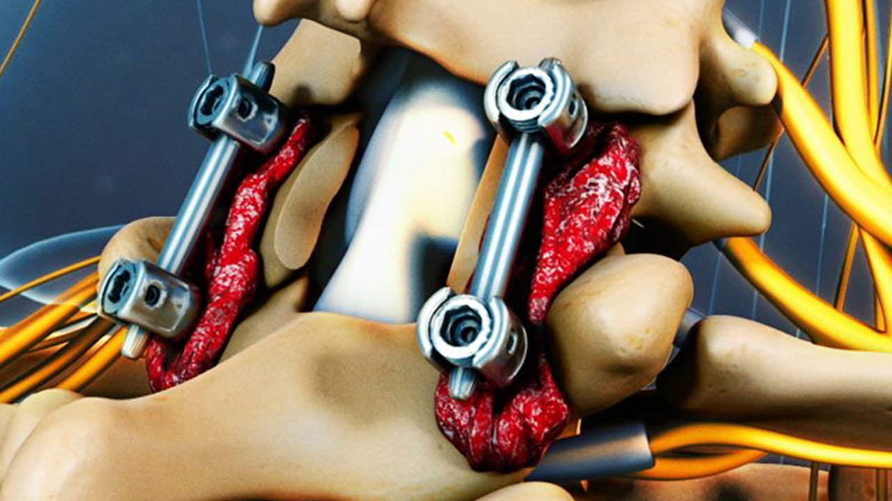
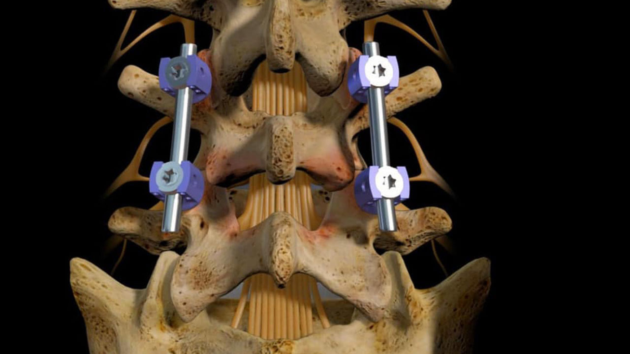
For patients with more serious spinal disorders, vertebrae fusion is required to immobilize the joints and stabilize the spine. Pedicle screws have been used to fuse vertebrae for a long time, but now this can be done with a “percutaneous” procedure. Instead of an open surgery, very small incisions are made in the skin. The surgeon uses X-ray fluoroscopic imaging to guide the placement of the special screws into the vertebrae.

The surgeon then attaches a rod to connect the pedicle screws, stabilizing the spine. This procedure is considered minimally invasive compared to traditional open surgery that splits and retracts the muscle of the spine through a much larger incision.

Schedule a Consultation
Spine Center Services